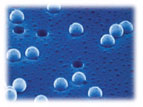
Synthetic and biological membranes and filters are widely used in a variety of industries and fields. The ability and efficiency of a membrane to remove target organisms, pathogens, or other particulates from a sample depends on pore size in addition to material properties of the membrane (e.g. charge, hydrophobicity, etc.).
Two primary methods exist for measuring the pore size and / or efficiency of the membrane, i.e. porometry and challenge testing. The former method involves application of a compressed gas to the pores of a filter that have been sealed with a wetting liquid. The gas pressure required to break the wet seal over the pores is correlated with pore size. To be truly quantitative, however, porometry requires cylindrical-shaped pores.1
Challenge testing is another method used to determine pore size that can accommodate membrane materials with various pore sizes and shapes. It also allows for material properties of the membrane and target particulate to play a role in determining the membrane’s filtration efficiency. Challenge testing involves the administration of one or more challenge particle types to a membrane followed by the analysis of particle size and abundance both upstream and downstream of the filter.
Challenge testing of synthetic and biological membranes and filters is used for a variety of applications such as:
- evaluating filtration efficiency of common commercially available filter classes (e.g. 0.2µm pore-size air filters, particle reduction filters, bioburden reduction filters, lab-grade and sterilizing-grade filters, etc.);
- aerosol challenge to test HEPA and ULPA filter integrity and in situ monitoring of cleanroom installation;
- evaluating integrity of synthetic barrier materials (e.g. latex gloves) to prevent viral-based disease transmission;
- evaluating fouling and biofilm formation on synthetic membranes; and
- evaluating media for filtration efficiency in removal of parasites or microorganisms from drinking water.
Filter challenge studies today often make use of highly uniform undyed, fluorescent, or visibly dyed polystyrene microspheres as surrogates for biologic or environmental particulates (e.g. microorganisms, pollens, dioctylpthalate esters (DOP), colloidal silica, A/C test dust, etc.) in order to reduce potential health risks and costs associated with the use of pathogens, atomized oils, etc.
Polystyrene is generally considered to be an inert polymer and it has a density of ~1.05 g/cm3 (i.e. slightly greater than that of water). Uniform polystyrene microspheres are available in a wide range of diameters (0.02µm – 10µm+) with a variety of surface modifications (e.g. non-functionalized, carboxyl-modified, amine-modified, etc.), and beads may be coated with protein or other molecules via straightforward methods to impart desired surface charge, hydrophilicity, or other properties. Visibly dyed and fluorescent versions of polystyrene microspheres feature encapsulated dyes, so the surface of dyed beads is also available for required modifications or coatings. A color palette of our visible dyes and excitation/emission spectra for our fluorescent dyes may be found in TechNote 103.
A variety of methods are employed for detecting polystyrene microspheres on filters, in the sample effluent, etc. Traditional methods for detecting undyed polystyrene microspheres include: microscopy (optical and electron), automated particle counters, turbidimetry and other light scattering-based techniques. Likewise, epifluorescence microscopy and fluorescence spectrophotometry have been widely used for detecting fluorescent polystyrene beads.
Methods such as flow cytometry, confocal laser scanning microscopy, and submicron particle counters are now also being employed for bead detection in membrane challenge testing. A number of articles citing use of newer and traditional bead detection methods may be found in the References section.2-9 Click to download Pdf version.
References
1. Rideal G. (2009) The Filtration Society: The importance of testing and standards. Filtration & Separation. Jan-Feb, 46(1):28-30.
2. Persson F, Langmark J, Heinicke G, Hedberg T, Tobiason J, Stenström TA, Hermansson M. (2005) Characterisation of the behavior of particles in biofilters for pre-treatment of drinking water. Water Res. Oct, 39(16):3791-800.
3. Spettmann D, Eppmann S, Flemming HC, Wingender J. (2007) Simultaneous visualization of biofouling, organic and inorganic particle fouling on separation membranes. Water Sci Technol, 55(8-9):207-10.
4. Finol EA, Siewiorek GM, Scotti CM, Wholey MH, Wholey MH. (2008) Wall Apposition Assessment and Performance Comparison of Distal Protection Filters. J Endovasc Ther, 15:177-85.
5. Ling TY, Wang J, Pui DYH. (2011) Measurement of filtration efficiency of Nucleopore filters challenged with polystyrene latex nanoparticles: experiments and modeling. J Nanopart Res, Oct, 13(10):5415-24.
6. Pall DB, Kirnbauer EA, Allen BT. (1980) Particulate Retention By Bacteria Retentive Membrane Filters. Colloids and Surfaces, 1:235-56.
7. Hou K, Gerba CP, Goyal SM, Zerda KS. (1980) Capture of Latex Beads, Bacteria, Endotoxin, and Viruses by Charge-Modified Filters. Appl Environ Microbiol, 40(5):892-6.
8. Zierdt CH. (1979) Adherence of Bacteria, Yeast, Blood Cells, and Latex Spheres to Large-Porosity Membrane Filters. Appl Environ Microbiol, Dec, 38(6):1166-72.
9. Widmer KW, Oshima KH, Pillai SD. (2002) Identification of Cryptosporidium parvum oocysts by an artificial neural network approach. Appl Environ Microbiol, 68(3):1115-1121.
10. Amburgey JE (2011) Removal of Cryptosporidium-sized polystyrene microspheres from swimming pool water with a sand filter with and without added perlite filter media. J Environ Eng, 137(12): 1205-1208.
Related Technical literature
TN 100 – Polymer Microspheres
TN 103 – Fluorescent & Dyed Microspheres
TN 203 – Washing Microspheres
TSD 726 – Decontaminating Microspheres
