Small Bead Kits for microvesicle and exosome analysis.
Posted: March 3, 2017
Interest in microvesicles and exosomes has rapidly risen over the past decade as a means to study cellular interactions, immune behavior, and various disease states. Flow cytometry, while traditionally focused on cell-based analysis, is a powerful tool being used to study these smaller microparticles. Due to the much smaller sizes however, analysis is not as straightforward. Researchers need to consider various limitations (such as the size sensitivity of their instrumentation) and make necessary adjustments.
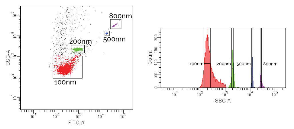
Both our Nanobead and Submicron Calibration kits are intended for use with small particle studies, allowing users to determine instrument capabilities, tailor acquisition settings, and make size estimations. The kits contain various sized fluorescent green nanospheres, with the Nanobead kit containing nominal 50 nm, and 100 nm beads (popular for exosome or viral analysis), and the Submicron kit comprising nominal 200, 500, and 800 nm beads (e.g. for microvesicles). The kits can be used together for a full complement of submicron sizes. Fluorescence offers an additional signal to detect and gate on, as traditional forward scatter acquisition is usually difficult, if not impractical.
Due to the heightened challenge in analyzing such small particles, we offer a tips and suggestions section in our Nanobead Product Data Sheet. Not all instruments will be able to see the smallest particles sizes and publications have often reported custom modifications. Examples of use, and additional product information are in PDS 834 and PDS 832.
References